


|
|
Leeper Group | ||||
| | |||||
|
|
|
|
|
|
|
  | ||||||
|
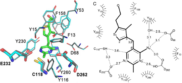
Abstract 146 Thiaminases, enzymes that cleave vitamin B1, are sporadically distributed among prokaryotes and eukaryotes. Thiaminase I enzymes catalyze the elimination of the thiazole ring moiety from thiamin through substitution of the methylene group with a nitrogenous base or sulfhydryl compound. In eukaryotic organisms, these enzymes are reported to have much higher molecular weights than their bacterial counterparts. A thiaminase I of the singlecelled amoeboflagellate Naegleria gruberi is the only eukaryotic thiaminase I to have been cloned, sequenced, and expressed. Here, we present the crystal structure of N. gruberi thiaminase I to a resolution of 2.8 Å, solved by isomorphous replacement and pseudo two-wavelength multiwavelength anomalous diffraction and refined to an R factor of 0.231 (Rfree, 0.265). This structure was used to solve the structure of the enzyme in complex with 3-deazathiamin, a noncleavable thiamin analog and enzyme inhibitor (2.7 Å; R, 0.233; Rfree, 0.267). These structures define the mode of thiamin binding to this class of thiaminases and indicate the involvement of Asp272 as the catalytic base. This enzyme is able to use thiamin as a substrate and is active with amines such as aniline and veratrylamine as well as sulfhydryl compounds such as L-cysteine and b-mercaptoethanol as cosubstrates. Despite significant differences in polypeptide sequence and length, we have shown that the N. gruberi thiaminase I is homologous in structure and activity to a previously characterized bacterial thiaminase I.
|
||||||
  | ||||||
 Cheryl A. Kreinbring, Stephen P. Remillard, Paul Hubbard, Heather R. Brodkin, Finian J. Leeper, Dan Hawksley, Elaine Y. Lai, Chandler Fulton, Gregory A. Petsko, and Dagmar Ringe,